CT EXAMPLE 01
Liver
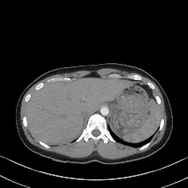
Reference label: Liver
Compare four real CT scans with their published anatomical annotations, then explore the illustrative chest radiographs below.
Explore the CT comparisons ↓X is the unmarked scan. Y is the same slice with a box enclosing the named structure in the published reference mask. These are anatomical references, not ParsecScope predictions or diagnoses.
Reference label: Liver
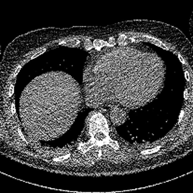
Reference label: Heart
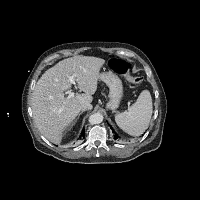
Reference label: Spleen
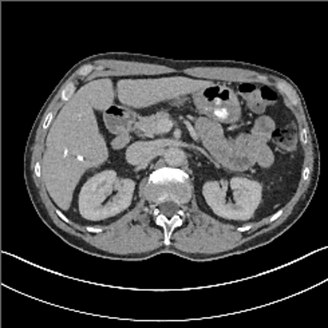
Reference label: Right kidney
CT scans and reference masks: Jakob Wasserthal, University Hospital Basel. TotalSegmentator, version 2.0.1, CC BY 4.0. Slices selected and displayed in grayscale; boxes added from the corresponding reference masks.
Explore a real chest radiograph alongside a marked view. The original image remains visible, so the meaning of an overlay is easier to follow.
These are real, publicly released medical images. The green review-region overlay was added for this presentation. It is not an automated finding, a medical annotation or a result from our service.
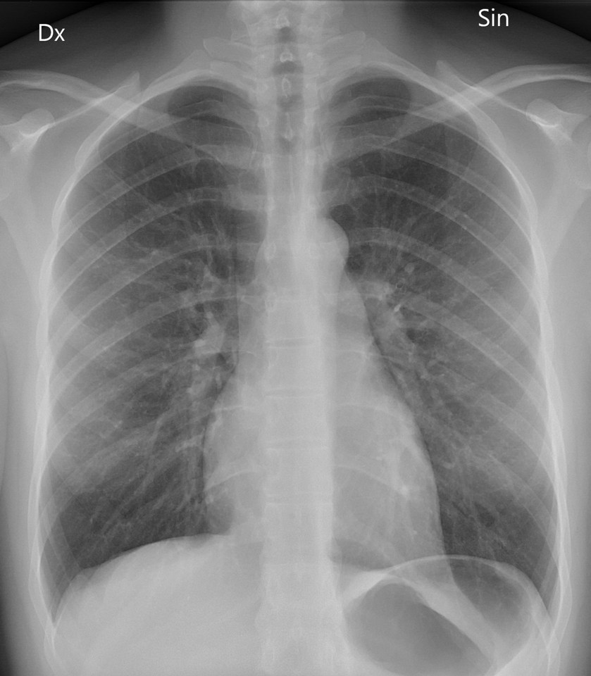 Original radiographDemonstration overlay
Side-by-side presentation helps viewers relate a highlighted region to its original surroundings.
A simple overlay explains the visual experience we are developing for medical image analysis and automatic marking.
Radiograph: Mikael Häggström / Wikimedia Commons · Original and CC0 terms. Resized for display; overlay added separately.
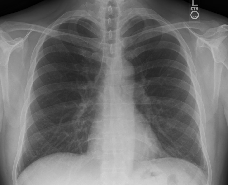 Original radiograph
Radiograph: Stillwaterising / Wikimedia Commons · Original and CC0 terms. Resized for display.